Overview
Level 1 autism (lower support needs) features an intact intellect with subtle but meaningful differences in how brain regions coordinate. A recurring pattern is local over-connectivity (stronger nearby links) paired with long-range under-connectivity (weaker distant integration). Combined with slightly lower synaptic density, this biases processing toward precise perception rather than broad contextual integration.
Specific Pathway Differences
Visual Pathways
Mildly enhanced occipital activation supports detail emphasis. Slight enlargement of the inferior fronto‑occipital fasciculus bolsters visuospatial skill. Under-connectivity with social networks may reduce effortless social scene integration.
Auditory Pathways
Sound–vision integration is modestly reduced. Subtle hypersensitivity can appear as smaller N1 amplitudes. Under-connectivity in superior temporal sulcus can mildly affect nuance parsing, while core speech discrimination stays intact.
Olfactory (Scent) Pathways
Mild hypoactivation of prefrontal regions during odor tasks; orbitofrontal variations may heighten sensitivity or aversion. Often linked to anxiety modulation rather than primary identification deficits.
Memory Pathways
Rote and detail memory can be slightly strengthened. A mildly enlarged hippocampus and increased theta coherence aid precise recall. Reduced alpha coherence may gently constrain integrative synthesis.
Cognitive Pathways
Perception & detail weighting outweighs broad executive integration. Under-connectivity in prefrontal (BA 9/10) can subtly influence flexibility, while local enhancements assist focused problem-solving.
Simulation Tie-In
The right (autistic) model reflects these dynamics: enhanced occipital responsiveness, cross-activation pathways, and increased stress/pain center sensitivity when Resources are low. Adjust sliders to observe how multi-sensory load plus resource depletion amplifies network spread versus targeted specialization.
Imagine your brain has a volume knob for every sense — sight, sound, smell, touch, and more. In a neurotypical brain, these knobs auto-adjust: they turn down background noise, dim awareness of scratchy clothing tags, and filter out the hum of fluorescent lights.
In an autistic brain, these knobs often stay turned up — or adjust unpredictably. When multiple senses are "loud" at once and your regulatory resources are low, the total sensory signal exceeds what the brain's filtering system can handle.
The insula — a brain region that bridges physical sensation and emotional experience — begins to process this overload as actual pain. This isn't imagined or "just anxiety" (though anxiety often follows). Brain scans show measurable activation in pain-processing regions during sensory overload. That's what this simulation demonstrates.
Sensory Hyperconnectivity (fMRI Evidence)
A 2025 study published in NeuroImage found significant differences in functional connectivity within the primary auditory and somatosensory regions in autistic individuals compared to neurotypical controls. Autistic brains showed heightened connectivity in these sensory areas — meaning sensory signals are amplified rather than filtered.
— Zhang et al. (2025), NeuroImage; abnormal FC of primary sensory network in ASD
A UCLA fMRI study on sensory overresponsivity (SOR) found that autistic individuals with high sensory sensitivity show stronger brain responses in both sensory processing areas AND the amygdala — the region responsible for threat detection and emotional reactions. This means the brain literally treats ordinary sensory input as threatening.
— Green et al. (2020), UCLA Health / Molecular Autism
Research on toddlers with autism found unusually strong connections between sensory areas of the brain, with stronger connections correlating with more pronounced autism traits. This suggests the hyperconnectivity pattern is present from very early in development.
— The Transmitter / Simons Foundation (2024)
Predictive Coding — Why Filtering Fails
A leading neuroscience framework called predictive coding helps explain why. Normally, the brain constantly predicts incoming sensory input and only "pays attention" to surprises (prediction errors). In autism, multiple theories describe how this system works differently:
- HIPPEA theory (Van de Cruys et al.): The autistic brain may assign inflexibly high importance to prediction errors — making unexpected sensory input feel overwhelmingly significant instead of being ignored.
- Hypo-priors hypothesis: Weaker prior expectations mean sensory input is experienced more "raw," with less modulation from past experience. Every signal feels fresh and intense.
- Atypical precision weighting (2023 computational models): Autistic individuals may struggle to adapt how much weight they give to changing sensory environments — unable to "turn down" processing when the environment becomes volatile or noisy.
— Van de Cruys et al.; Pellicano & Burr (2012); Lawson et al. (2023), Frontiers in Psychiatry / PLoS Comp. Bio.
The Insula & Pain Processing
A meta-analysis found consistent anterior insula hypoactivity in autism — meaning this critical integration hub doesn't function typically. The insula normally helps you distinguish "this is just a scratchy tag" from "this is a wound." With altered connectivity, it may process ordinary sensory overload as genuine pain signals.
— Uddin & Menon (2009); Di Martino et al., Biological Psychiatry meta-analysis
A 2024 study found that altered dynamic functional connectivity of insular subregions can predict symptom severity in autistic individuals — directly linking insula wiring patterns to lived experience.
— Wang et al. (2024), Semantic Scholar
fMRI studies show greater activation in the anterior cingulate cortex during pain anticipation in autistic adults, and autistic adults report significantly higher rates of chronic pain conditions. Importantly, the posterior cingulate and insula showed increased activation specifically when experiencing unpleasant stimuli.
— Failla et al.; Duerden et al., PAIN / J. Autism Dev. Disord.
Weaker Neural Suppression (Why It Won't Turn Off)
A University of Washington fMRI study found weaker neural suppression in the visual cortex of autistic individuals — meaning the brain is less effective at dampening repeated or irrelevant visual signals. This helps explain why a flickering fluorescent light that a neurotypical person stops noticing after seconds can remain intensely distressing for an autistic person.
— Murray et al., University of Washington / Current Biology
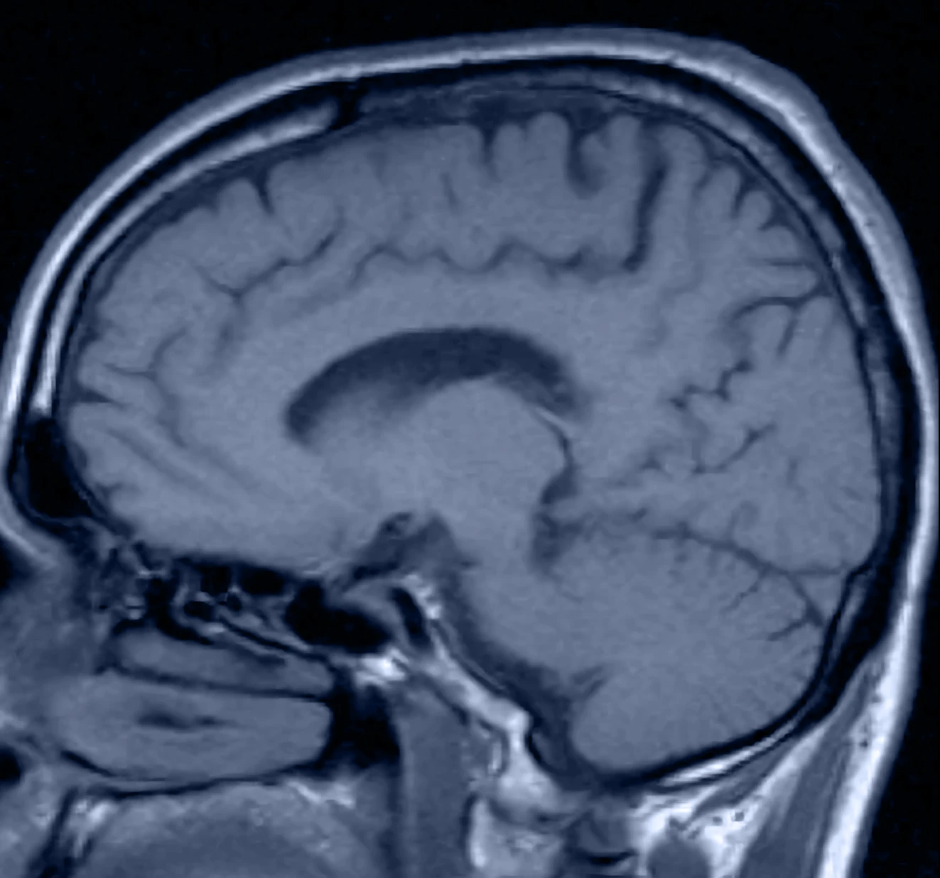
White Matter Differences (Structural Evidence)
A February 2025 study in NeuroImage using diffusion-weighted imaging found significant differences in white matter fiber tracts in young children with ASD, especially in the left hemisphere. These altered pathways showed impaired integrity and developmental delays in critical brain networks — providing a structural basis for the functional connectivity differences observed in fMRI.
— NeuroImage (Feb 2025), News-Medical.net summary